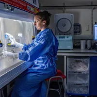
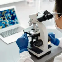
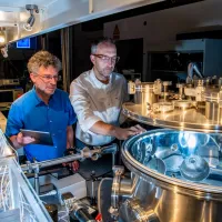
Our 3D X-ray histology (XRH) facility is a collaboration between the µ-VIS X-ray Imaging Centre and the Biomedical Imaging Unit and funded by Wellcome Trust.
We provide micro-computed tomography (micro-CT) imaging of soft-tissue samples integrated with a workflow which includes reliable sample traceability, correlative imaging visualisation, data sharing and reporting, and automatic metadata parsing.
Advantages of XRH
Micro-CT has several advantages over traditional histology:
ability to visualise and quantify microstructure soft tissue in its 3D context
non-destructive and can be combined with specific 2D histology techniques, including histochemistry, immunocytochemistry and in situ hybridisation
ability to image archival material stored in hospitals and tissue banks to allow validation of microstructural hallmarks of disease
coupled with artificial intelligence/computer-aided diagnosis it improves diagnostic accuracy and support patient stratification
Our services
We welcome collaboration proposals from scientists who want to explore and discover the potential of XRH for their research portfolio. Our services include:
production of XRH images
image analysis involving sample visualisation and measurement
correlative imaging,combining XRH with conventional histology, immunohistochemistry, and/or other microscopy techniques such as serial block face SEM)
bespoke analysis, such as, image based model generation (meshing) for FE/CFD modelling or 3D printing
MILabs U-CT 3D/4D optical imaging and microCT system
This scanner can scan living organisms up to the size of a small rabbit (up to five mice can be scanned at once). It features:
correlative imaging to map fluorescence/luminescence images onto X-ray CT images and white light images
data manipulation via MiLabs software and/or third party software
µCT modality involving spatial resolution down to c. 13 μm, 3D fluorescence, bioluminescence and Cherenkov luminescence tomography
Optical system involving resolution of c. 3 mm, visual to near-IR range (340 nm to 830 nm, up to six emission filters and up to 11 excitation filters and a C-MOS detector
Nikon Med-X (alpha)
This was designed as a soft tissue imaging research tool for biology and medicine.
130 kVp X-Ray source
optimised for biomedical imaging & unstained FFPE histological specimens
standard reflection target (~2 µm spot)
2 x 2k flat panel detector
standard cone-beam & helical scanning
samples up to ~300 mm and up to 15 kg
Nikon XTH225ST XRH 3D X-ray histology scanner
This can be used for non-destructive radiographical and tomographical inspection of biomedically relevant specimens, and is optimised for high-throughput imaging of unstained formalin-fixed and paraffin-embedded (FFPE) soft tissue specimens.
Nikon multi-target micro focus X-ray source
130 kVp / 130 W Mo rotating target (custom)
10 μm spot up to 50 W
180 kVp / 20 W Mo transmission targets (custom)
1.5 μm spot up to 5 W
225 kVp / 225 W multimetal reflection target
2.5 μm spot up to 7 W
sample autoloader | up to 10 specimens per rack (custom)
optimised for high throughput 3D X-ray Histology
2.8 x 2.8 k 16-bit flat panel detector
standard cone-beam & helical scanning
samples up to ~400 mm and up to 50 kg
SkyScan 1176 in-vivo CT scanner
This scanner allows full body mouse and rat scanning or distal limb scanning for larger animals, such as rabbits. It also provides scanning flexibility to allow imaging of a wide range of samples from lung tissue to bone with titanium implants.
small animal pre-clinical research
100 kVp reflection source
11MP camera
9µm in vivo spatial resolutions achievable
integrated physiological monitoring
bone morphometry tools / 2D & 3D quantitative analysis / 3D visualization
Research
We have designed our XRHworkflow to fit seamlessly into current histology workflows in biomedical and pre-clinical research, as well as clinical histopathology.
Research applications
head and neck tumour visualisation
assessing the effectiveness of cancer treatments
understanding the biological response to cochlear implantation
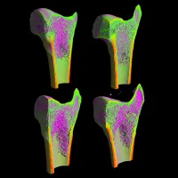
imaging of the bone vascular network to study osteoporosis.
Expert team
The XRH team is made up of experienced X-ray beam scientists, microscopists, biomedicalresearchers and pathologists, together with computing and information scientists. The team can support and develop a broad range of biomedical imaging applications.